
A Patient's Guide to Laser Correction Surgery: Steps, Risks and Requirements
- Atanas Bogoev M.D.
- Oct 16, 2023
- 11 min read
Updated: Sep 22, 2024
If you have concerns about wearing glasses or contact lenses to have clear vision this article is just for you. Laser correction surgery is a practical way to cut your daily dependence on glasses or contacts.
Are you considering laser vision correction? The ultimate decision of whether to undergo such a procedure depends on YOU. This Ophthalmology24 publication is not meant to assist you in making a decision. The goal is to provide you with all the information about laser eye surgery in one place.
Laser correction surgery has been performed for many years using excimer lasers. Nowadays, in some of the techniques, in addition to the excimer laser, eye doctors also use a femtosecond laser. We will tell you about the difference later in the article.
The choice of the right technique for the best results depends primarily on the parameters of your eye. But also on your profession, your hobbies, and personal preferences.
Before heading for laser surgery, discuss your preferred choice of laser correction surgery with the refractive surgeon. That's not only imperative from a medical point of view but also from a lifestyle perspective.
How Does Vision Work?

Light rays enter the eye through the front surface - cornea, pass through the pupil, and go through the intraocular lens.
The cornea and the lens are responsible for focusing the rays onto the retina. About 2/3 of the eye's focusing power comes from the cornea, and 1/3 comes from the lens. The retina converts the light rays into impulses, sending them to the brain through the optic nerve.
In the brain (visual cortex), these impulses are perceived as images.
RELATED: What Can Damage Your Eyesight?
Causes of Unclear Vision
1. Nearsightedness (Myopia)
A condition in which nearby objects appear clear, but distant ones appear blurry. Myopia is most often due to an eye with a longer length, which leads to the focal point of light rays being in front of the retina.
2. Farsightedness (Hypermetropia/Hyperopia)
We associate hypermetropia with unclear vision, eye strain, and headaches (usually after intense reading or computer work). In farsightedness, the eye typically has a shorter length, causing light rays to focus behind the retina.

3. Presbyopia (Age-Related Farsightedness)
Age-related farsightedness is an inevitable consequence of aging. It begins to affect most people around the age of 40.
Starting from around that age, people (who have had no trouble with distant vision) usually need the help of reading glasses. Individuals who have already needed distance glasses before this age often require two pairs of glasses (or one pair with progressive lenses for near and far correction).
The first complaints of presbyopia arise when reading very small print (e.g., medical leaflets), especially in poor lighting. To compensate, people usually hold the text further away to achieve a clearer vision. Over time, their dependence on reading glasses increases.
4. Astigmatism
In the condition astigmatism, simply said, the shape of the cornea is not a perfect sphere, but rather ellipsoid. This causes focused light to have many different focal points in the eye, leading to unclear or distorted vision. Patients with astigmatism often have either myopia or hypermetropia as well.
RELATED: Life with Astigmatism
5. Irregular Corneal Surface (High-Order Aberrations)
Sometimes issues with blurry vision are more complex than those described above.
This typically happens when the surface of the cornea has an irregular shape (multiple fine random irregularities). Aberrations (distortions) can be due to previous corneal surgery, injury, or other eye conditions.

Requirements for Laser Correction Surgery
Not everyone is suitable for refractive surgery. The final assessment of whether you are a good candidate depends on the eye doctor (refractive surgeon).
There are strict criteria to meet to minimize complications and achieve the most lasting result:
The minimum age for correction is 18 years old
You should NOT be pregnant or breastfeeding
You should NOT have corneal disease
The prescription of your glasses or contact lenses should have been stable for at least 1 year
You should be well informed about the potential risks associated with different types of procedures (see below)
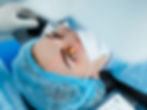
Laser Eye Surgery Principles

Laser correction uses an Excimer Laser that changes the shape of the cornea and modifies its refractive power. In simple words, the laser makes the cornea either more or less powerful in bending light.
The laser beams are typically very precisely directed using a 6-D tracking system that scans and tracks eye movements over 1000 times per second. Additionally, a special system ensures the proper alignment of the laser beams with the size of the pupil, and the laser is adjusted as needed.

Laser Treatment for Myopia
In the treatment of myopia, the surgeon uses the laser to remove a central circular area of corneal tissue. Thus making the cornea flatter and reducing the eye's refractive power. The tissue is removed in a complex manner that is set into the laser's computer by the surgeon. When myopia is too high or the cornea is too thin, laser correction is not possible.

Laser Treatment for Hyperopia
In the treatment of hyperopia, the surgeon uses a laser to remove tissue from the periphery of the cornea (similar to a small crescent). Thereby making the central cornea steeper and increasing its refractive power.

Laser Treatment for Astigmatism
In the treatment of astigmatism, the laser selectively removes corneal tissue in the shape of an ellipse to achieve a smooth and symmetrical cornea. Imagine changing the shape of a rugby ball into a spherical basketball.

Laser Treatments for Other Conditions
To treat high-order corneal aberrations, doctors use special data from an individual corneal map (topography). It is always approached on an individual basis - custom laser correction surgery. This is used in addition to correcting the other refractive abnormalities, and rarely as a standalone indication for laser refractive surgery.
Types of Laser Vision Correction
We can divide laser correction surgery into several major categories: ReLEx, LASIK, and Surface Methods for Laser Correction.
In ReLEx part of the inner layer of the cornea is cut with a laser and removed.
In LASIK, a partial section of the cornea (flap) is partially cut and lifted, and the actual reshaping of the cornea is done under this flap.
While in surface methods, doctors perform the entire procedure directly on the corneal surface.
LASIK
With LASIK, vision restoration is quick. Patients can usually return to work and drive after about 1-2 days. There is only slight discomfort for a few hours after the operation. Patients usually have follow-up appointments a few days after the procedure, at 1 month, and at 3 months.
Video Credit: Vold Vision
ReLEx (SMILE)
Another modification of laser corrective surgery is called ReLEx (Refractive Lenticule Extraction) or SMILE , in which a small cut of the middle of the cornea is performed with a femtosecond laser, and later removed completely through a small incision (also performed by the same laser).
The advantages of ReLex are like those of LASIK and result in faster post-operative recovery. Yet, the correction with a femtosecond laser does not offer other advantages in terms of visual quality after the procedure.
Video Credit: Clinic New Vision
Surface Methods for Laser Correction
The surface methods are:
PRK
LASEK
Epi-LASIK
Trans PRK
Using this method, the epithelium (the outermost layer of the cornea, which regenerates after a few days) is removed. Next, with the excimer laser, similar to LASIK, the refractive surgeon reshapes the corneal stroma (inner layer of the cornea). Finally, the doctor places a soft contact lens without a prescription on the cornea, acting as a bandage. The contact lens remains in the eye for 4-5 days until the corneal epithelium heals.
Surface methods are initially more uncomfortable than LASIK. Yet, they are more suitable for patients with thin corneas and those participating in sports, military, or extreme sports.
In surface methods, vision recovers slowly, which means you won't be able to return to work for the next 5-7 days. Typically, your vision won't be good enough to drive for 7-10 days after the correction.
The vision will continue to vary within the first month after the operation, being clearer on some days and blurrier on others. For some patients, achieving clear vision can take more than a month.
Please note that the eyes may be uncomfortably painful and very sensitive to light for about 5-7 days.
Patients are usually scheduled for a follow-up appointment 4-5 days after the correction. Then at 1 and 3 months.
Now, let's talk about the only difference between the various types of surface methods. That's the way the surface layer of the cornea (the epithelium) is removed.
PRK
In PRK (PhotoRefractive Keratectomy), the epithelium is mechanically removed by the surgeon with a special blade under local anesthesia (eye drops).
Video Credit: ΟΦΘΑΛΜΙΑΤΡΟΣ ΕΥΟΣΜΟΣ - ΜΠΙΚΟΣ ΚΩΝΣΤΑΝΤΙΝΟΣ
LASEK
In LASEK (Laser Sub-Epithelial Keratectomy), the corneal epithelium is removed by the surgeon using a special alcohol solution and a blunt instrument. The alcohol solution makes the removal of the epithelium less traumatic compared to PRK.
Video Credit: EyeSmart
Epi-LASIK
In Epi-LASIK, the epithelium is removed by the surgeon using a special machine under the surgeon's control.
Trans-PRK
In the newest surface method, Trans-PRK (tPRK), the epithelium is entirely removed by the laser and does not come into contact with the surgeon. Trans-PRK is a new and quite convenient technique but is not suitable for all patients. The determination of the best correction method is made by the treating ophthalmologist.
Which is better: RELEX, LASIK or Surface Methods?
Simply said - there is no clear answer. Each method has its own upsides and downsides.
The best option depends on the patient's age, type of refractive error, thickness of the corneal tissue, personal lifestyle, and expectations.
Discuss with an eye doctor the most suitable method for each individual case.
RELEX/SMILE (Refractive Lenticule Extraction):
The advantages are it preserves the stability of the cornea, while not lasering the the corneal surface itself, thus reducing the risk of dry eyes, and allowing for faster recovery.
The disadvantages are the additional financial expenses, compared to other methods, not being able to correct high degrees of astigmatism or farsightedness, and it is not reversible or adjustable.
LASIK (Laser-Assisted In Situ Keratomileusis):
The advantages of LASIK are it can treat a wide range of myopia, hyperopia, astigmatism, and high-order aberration errors while providing rapid visual recovery and minimal discomfort. LASIK is usually reversible and adjustable in case of under-correction or over-correction.
The disadvantage is it involves cutting the corneal surface, which increases the risk of complications such as dry eyes, LASIK flap displacement, or infection by contamination.
Surface Methods:
The benefits are they preserve the strength and stability of the cornea, eliminate the risk of flap-related complications (because no flap is created), and are suitable for patients with thin or irregular corneas.
The drawbacks are they cause more pain and discomfort after the procedure, require longer healing time and the visual recovery is gradual (fluctuating in the first few days). The surface methods may sometimes cause scarring or haze (fogging) of the cornea.
In summary about ReLEx (SMILE) correction
A thin strip of the middle of the cornea is cut with the laser and carefully removed
Rapid healing
Minimal discomfort
In summary about LASIK laser correction surgery
A "flap" is cut from the cornea
Rapid healing
Minimal discomfort
LASIK can be performed with a microkeratome or femtosecond laser (nowadays usually almost entirely with femtosecond laser)
In summary about surface methods - PRK, LASEK, Epi-LASIK, Trans PRK
No "flap" is cut from the cornea
longer time for healing is required
More discomfort post-intervention
Suitable for thin corneas
Laser Vision Correction Step by Step
Here is the typical timeline for having laser correction surgery:
1. Motivation to stop wearing glasses/contact lenses
2. Initial eye examination to prepare for laser vision correction (3-4 hours)
Eye exam to determine the actual prescription with pupil dilation
Interpretation of results by the refractive surgeon
Determination of the correction method
Setting a date for the laser refractive surgery
3. Show up on the day of laser eye surgery (2-3 hours)
Pre-procedure examination - checking optical correction again (20-30 minutes)
Performing the actual laser correction (15-20 minutes)
Stay at the clinic (30 minutes)
Eye doctor provides instructions for post-operative care (eye drops, rest, scheduling a follow-up)
4. Follow-up examination 4-7 days after the correction (10-20 minutes)
Laser Correction Surgery Risks
This may be one of the few articles where all the risks of the procedure are openly addressed. The goal is not to discourage you from correcting your vision. But on the contrary - we believe the patients should be as informed as much as possible about the decisions they make.

Like with any surgery, complications during RELEX (SMILE), LASIK, and surface methods for laser correction are possible. Fortunately, they are quite rare.
Insufficient correction, over-correction, or the development of astigmatism can occur, but this can usually be improved with glasses, contact lenses, or additional laser correction surgery.
Most complications are treatable without leading to permanent vision loss. Then again, there's a rare chance your vision may not be as good after the surgery as it was before, even with glasses or contact lenses.
Almost every patient experiences some level of dryness and variable vision during the recovery period. For most people, these symptoms go away within a month. However, some patients may continue to experience symptoms for a longer time.
Other adverse reactions and discomforts for several weeks after refractive surgery are:
Redness
Scratchy sensation
Glare
Halos
Starbursts around lights
Light sensitivity
Most of these adverse reactions dissipate over time, but in rare cases, they can be permanent.
Infections after laser correction surgery are possible but are usually well-treated with antibiotics. Complications related to the flap in LASIK are rare and may need additional surgery. In surface methods, there's a rare chance of haze developing at the center of the cornea, which may require further surgery.
Rarely, even after laser refractive surgery, some people may still need to wear glasses or contact lenses.
Still need glasses after laser eye surgery?
The goal of any laser correction is to eliminate the need for glasses. However, there is a rare possibility of the prescription returning. This most commonly occurs in individuals under the age of 20 whose prescription for glasses progresses before undergoing the procedure.
Laser correction surgery doesn't stop the growth or changes in your prescription; it simply eliminates your current prescription.
This is why the examination before laser intervention and the experience of the refractive surgeon is of utmost importance.
Even if this happens, in most cases, refractive surgeons can perform another refractive surgery procedure to remove any residual or newly developed refractive error (diopter change).
Laser Correction for Presbyopia
None of the standard methods for refractive surgery can prevent the development of presbyopia (age-related farsightedness).
Most people over the age of 40 with excellent distance vision end up needing reading glasses, whether they have had laser correction surgery or not.
Nonetheless, there are options for what's called Presby LASIK (Multifocal LASIK, Bifocal LASIK, and Monovision LASIK), aiming to leave one eye nearsighted (for good near vision) and the other with good distance vision.
In addition, the intraocular lenses in people over 60 undergo age-related changes, which can further worsen the effects of correction.
RELATED: Eye Conditions from Old Age
Preparing for a Laser Correction Pre-Examination
After scheduling an appointment with an ophthalmology specialist, you need to prepare as follows:
Stop wearing your contact lenses before the initial eye examination. 1 week for soft contact lenses and 2-3 weeks for rigid and rigid gas-permeable contact lenses. Otherwise, the doctor won't be able to make the most accurate assessment, and a repeat visit may be necessary.
Avoid putting on eye makeup. It could interfere with the proper imaging of the corneal maps and might lead to missing some details.
When scheduling an appointment with a refractive surgeon for laser correction, take all your medical history documents. Especially past glasses prescriptions, eyedrops (if you are using any), and current glasses.
During the initial exam, you will probably receive eyedrops for pupil dilation. Dilating the pupils helps the ophthalmologist examine your eyes better before scheduling laser correction surgery.
You will not be able to drive as everything will be blurry. You will also be light-sensitive, so having a pair of sunglasses can be helpful.
If your myopia (nearsightedness) is constantly worsening, you may not be a suitable candidate for laser correction. Still, you should have a consultation with an eye doctor for your eye health.
Learn more about eye care in the Ophthalmology24 blog.
Resources:
✅ Medically reviewed
Checked by Atanas Bogoev, MD.